
I’m sure most of us are familiar with reading a telephone number only to forget a few of the numbers when punching it in the phone, or burning the toast while simultaneously cooking eggs and talking to a friend. All of these are lapses in working memory, which is crucial for organizing one’s day as well as planning and carrying out a sequence of events.
What is Working Memory?
Working memory is the brain system that holds information for limited time intervals while we are making decisions or performing calculations. Simply, it is keeping in mind everything that is required to keep in mind while performing a task. It is critical for a wide variety of tasks, from doing arithmetic and temporarily memorizing phone numbers to navigating a new environment and negotiating spatial obstacles.
The term was first used by George Miller and colleagues in 1960. Around the same time, Miller coined the magic number 7, plus or minus two, which refers to the now-refined number of objects an average human can hold in short-term memory. The term was then elaborated on by Alan Baddeley who described working memory as a form of short-term memory that integrates moment-to-moment percepts over a short time period. These perceptions are then related to memories of past experiences in order to execute planning and complex behavior.
Baddeley developed a model of working memory, known as the multi-component model, which has proven useful and resilient in applied psychology and neuroscience over the decades. He envisioned working memory not as a single storage cache, but as two short-term memory buffers, one for verbal, which he called the phonological loop, and one for visual memories called the visuospatial sketchpad. The phonological loop is a cache for speechlike memory traces that are registered and decay within seconds unless rehearsed either verbally or subvocally. Generally, recall declines as both list length and word length increase, reflecting Miller’s law.[1] The visuospatial sketchpad is a subcomponent that stores or processes information in a spatial or visual form and is utilized in tasks such as spatial navigation.
Managing both of these is the central executive, an attentional control system that manipulates and coordinates information stored in the buffers for problem-solving and planning. Later, a fourth subsystem was added on, the episodic buffer, which is seen as a temporary storage area of episodic chunks formed from the other two subsystems that interfaces with past experience and/or consolidates the information into long-term memory.
While long-term memory stays with us even when we’re not actively retrieving them, working memory is an executive function that works more like an active process, a short-term mental workspace where we maintain and manipulate the information we need at a given time. However, working memory and long-term memory are both closely interrelated, as seen by numerous imaging studies where long-term memory centers such as the hippocampus activate concurrently with working memory areas. Additionally, researchers find increased recall in words that form meaningful sentences, reflecting the importance of long-term memory features such as grammar, syntax, and meaning in working memory tasks.
What Brain Regions are Involved in Working Memory?
After Baddeley defined his working memory model, Joaquin Fuster at the University of California, Los Angeles and Goldman-Rakic linked the prefrontal cortex (PFC) to Baddley’s studies on working memory. They found that removing the PFC does not result in a general deficit of short-term memory, but rather a deficit in the functions that Baddeley described as working memory. The prefrontal cortex is a part of the frontal lobe that mediates complex mental processes such as organizing, planning, learning, cognitive flexibility, and memory.[2]
Executive functions were commonly thought to occur in just primates, given that these complex mental processes are associated with the “higher order” cognitive functions stemming from the more complex primate cortex. However, the rodent medial prefrontal cortex shares strong anatomical homology with primate PFC, particularly the dorsolateral prefrontal cortex. Studies have elucidated that the medial prefrontal cortex is critical in working memory for both spatial and visual object information, with the amount of prefrontal activation dependent on the amount of information held in working memory.[3]
The PFC across species has been shown to utilize a division of labor system under certain test conditions, with parallel processing of working memory features such as location, object, or words. That is to say, working memory can be divided at the level of PFC depending on the sensory modality processed as the cortex cooperates with connected sensory regions that hold and use the information for brief moments of time. For instance, Goldman-Rakic and colleagues found that one part of the PFC is involved in working memory for object identity and another for spatial locations.[2]
One factor that heavily influences this processing scheme is the delay period before working memory is assessed in the choice period. If the delay period is long enough, the PFC has been purported to work in conjunction with the hippocampus as an integrated circuit, coordinating retrospective and prospective memory processes in achieving the desired goal.[3] This finding has been supported by lesioning studies in rats and mice, where researchers who inactivate the hippocampal formation are able to impair working memory performance in the radial maze. The area is purportedly important for the acquisition and maintenance of spatial information in regards to the location of the baited arms, and/or the reference to episodic memories when navigating the maze.
Development of Working Memory in Humans and Rodents
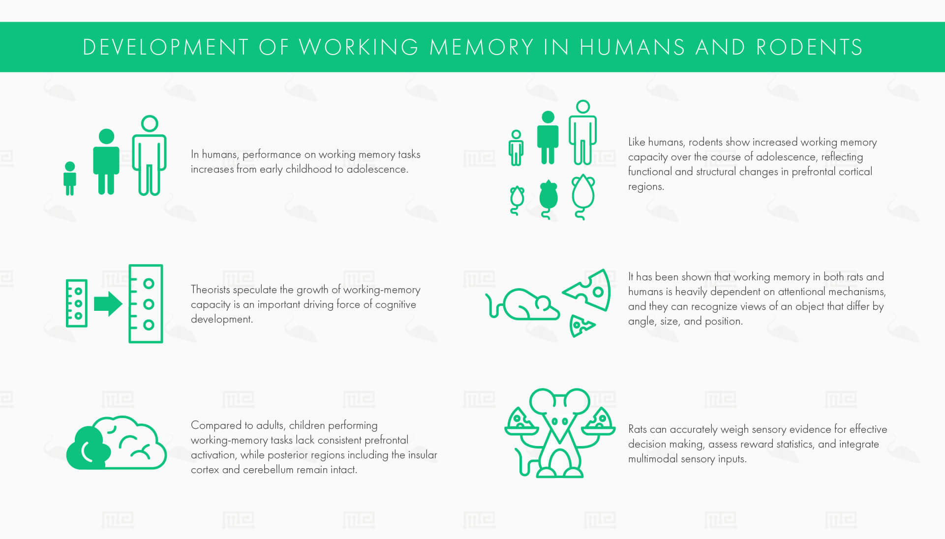
In humans, performance on working memory tasks increases from early childhood to adolescence. Theorists speculate the growth of working-memory capacity is an important driving force of cognitive development, as the capacity of working memory strongly predicts cognitive abilities in childhood and capacity at one age can accurately predict reasoning ability at a later age. Compared to adults, children performing working-memory tasks lack consistent prefrontal activation, while posterior regions including the insular cortex and cerebellum remain intact.
Like humans, rodents show increased working memory capacity over the course of adolescence, reflecting functional and structural changes in prefrontal cortical regions. Adolescent rats show reduced working memory performance until postnatal day 50, by which time performance is not significantly reduced compared to adults.[4] Evolutionarily, working memory holds a common essential use in humans and rodents for recognizing the spatial location, avoiding spatial obstacles, and organizing general goal-directed behavior for survival.[3]
Rat working memory shares many functional characteristics with humans. It has been shown that their working memory is heavily dependent on attentional mechanisms like humans, and they can recognize views of an object that differ by angle, size, and position. They can also accurately weigh sensory evidence for effective decision making, assess reward statistics, and integrate multimodal sensory inputs. Humans may be unique in their utilization of inner speech (such as for verbal rehearsal) and the frequency of use and high flexibility of task-independent working memory abilities.[3][5][6]
Behavioral Assays for Assessing Working Memory
The crucial neuroanatomical and functional characteristics of the human PFC are largely preserved in rodents, making cross-species comparisons and translational research findings meaningful and effective.[1] As such. rodent models are promising tools in advancing our understanding of age-related executive function decline and the impact of disease and other cognitive disorders on working memory. In addition to various rodent models, we will also take a look at a few of the popular working memory assays in non-human primates and humans.
Behaviorally, working memory is activated when tasks involve:
- different stimuli which govern the criterion response across different trials
- a cue the subject must remember which varies from trial to trial
In the tasks described below, episodic memories are elicited by both visual and spatial cues that guide the rodent’s goal-directed behavior. The task-related information is encoded by prefrontal areas and corresponds to working memory across variable delay periods. In nearly every case, lesions in this area affect the ability to retain this memory.
This is in contrast to reference memory, which is mostly governed by the hippocampus and is known to remain mostly constant over time. However, as mentioned, co-activity between the regions associated with working memory (PFC) and the hippocampus is commonly described depending on the task design.[7]
With humans, working memory includes both recall and manipulating the information being remembered (e.g. adding two to a string of digits or reversing a number sequence). In rodents, this scheme is generally infeasible, so increasing the delay duration is often used as a method of increasing working memory load and noting spatial working memory performance across variable inter-trial intervals.
The Delayed Alternation Task
In the delayed alternation task, either a T-maze or a Y-maze is utilized in a binary choice paradigm. This assay exploits the rodent’s natural preference for novelty (termed spontaneous alternation). Rats are first placed on the stem of the maze and rewarded for entering either of the arms. Then, after a delay, the rat is rewarded only if they entered the arm not chosen on the previous trial. Often, the correct choice alternates between the left and right arms across trials. Working memory is assessed by varying the inter-trial interval, usually between 10 to 60 seconds, and noting the number of correct choices. Lesioning the medial PFC impairs performance in both spontaneous and learned alternation, supporting its role in working memory.[5]
The Radial Arm Maze
The radial arm maze has been used for over four decades and involves a similar information trial and retention trial format. In the information trial, the researcher places rewards on 4 of the radial arms, with the other 4 arms blocked. The rat begins in the center platform and promptly visits each open arm to obtain the rewards. Following a delay period, the rats in the retention trial navigate the maze with all the arms open, but only the blocked arms from trial 1 are baited. Rats must recall trial-unique information to maximize the reward, namely, to avoid the rewarded arms during trial 1 and avoid re-visiting arms baited during trial 2 when the reward is collected.[5][8]
The Morris Water Maze
The standard Morris water mazes can be used to assess working memory in addition to land-based tasks, providing advantages such as no food restrictions and rapid learning. In the delayed match-to-place version of the water maze, there is similarly an information trial and retention trial. In the former, the rodent must find a hidden submerged platform. After a variable delay, a retention trial is then conducted, with the platform in the same location. The rats are required to remember trial-unique information, i.e. the platform location, across the delay period, which ranges anywhere from thirty minutes to six hours. This location is changed each day and the memory for the location is assessed using a difference measure. Since the platform location must be differentiated from the previous locations in memory, this task strongly involves PFC activity. This maze tests spatial and working memory by observing and noting variables such as escape latency, thigmotaxis duration, distance moved, and velocity during the time spent in the water maze tank.[8]
The Barnes Maze
Working memory can also be delineated with the Barnes maze, which shares similarities to the radial arm maze. In this task, there is a well-lit circular central platform with holes around the edges, and only one hole leading to an escape tunnel. There is no food reward in this case, as the motivation arises from the desire to escape the brightly lit area. Accurate performance requires the rodent to learn and remember the correct hole location, indicating both spatial regions of the brain and the hippocampus are involved in this assessment. Working memory errors are quantified by revisits to incorrect holes that have already been investigated in the trial.[5]
The Spatial Delayed Response Task
A classic working memory assessment in non-human primates is the spatial delayed response task. Information regarding spatial location (the location of the reward in one of two food wells) must be held over some delay interval and accurately recalled in a choice period. An analogous operant procedure was developed in rodents using levers, where the rats press a lever in a starting phase which operates a light over the food trough, then after a delay of variable length, the rat must press the same lever to obtain the reward. Dorsolateral PFC lesions disrupt performance on these spatial delayed response tasks. Electrophysiological recording from this area shows persistent spatial tuning during the delay period of these tasks, confirming its critical role.[5]
Assessing Working Memory in Humans
In humans, working memory and working memory capacity is assessed with various neuropsychological measures, one of the most common of such is the working memory complex span tests. In digit spans, the subjects must recall the correct sequence of digits that is heard or seen, while in reading spans, the subjects must read a series of unconnected sentences aloud to remember the final word of each sentence of a series.
The Effects of Aging on Working Memory
In humans, working memory is believed to increase gradually over childhood and declines gradually in old age.[6] According to some theories, the slower processing speeds associated with age allows more time for working-memory contents to decay, reducing total working memory capacity. An alternate, empirically-supported hypothesis states that deficits in working memory due to old age arise from the inability to inhibit irrelevant or distracting information. This leads to working memory being cluttered with irrelevant contents that reduce the effective working memory capacity. One neurobiological explanation put forward by Robert West at DePauw University is that the prefrontal cortex shows the highest degree of age-related atrophy. Synaptic density similarly declines in the frontal lobes in a preferential way with age.[6]
This finding is also noted in aged rodents across the operant tasks described above. Many investigators have noted delay-dependent impairments in aged rats and mice on delayed alternation tasks in comparison to the younger subjects. While the aged rats’ performance was similar to the young cohorts on short delays, the deficits were evident by an increase in the number of errors at long delay periods.[5][6]
Conditions and Disorders that Affect Working Memory
ADHD
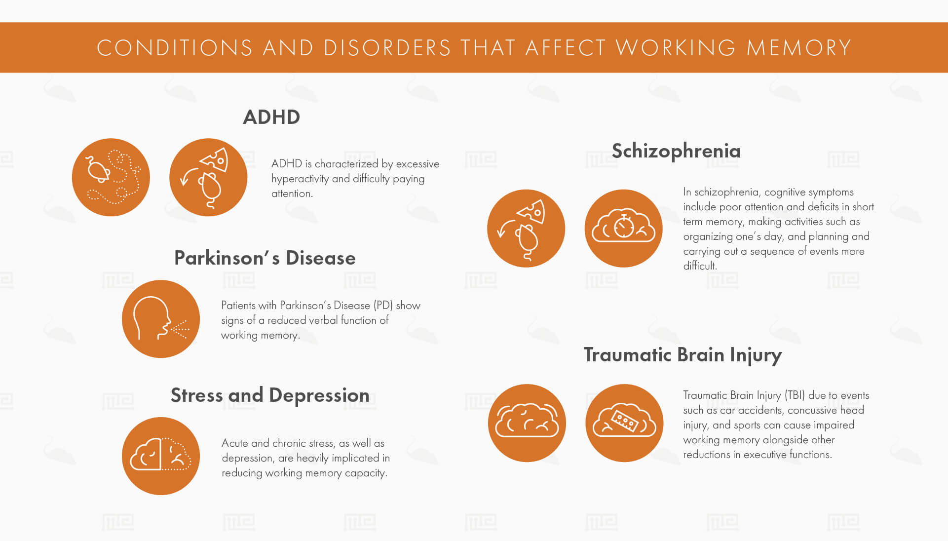
ADHD is characterized by excessive hyperactivity and difficulty paying attention. Several studies have found deficits in visuospatial and verbal working memory, as well as other executive function tasks such as response inhibition.[9] The neurobiological underpinnings of ADHD are thought to involve frontostriatal dysfunction, particularly disruption of the dopaminergic and noradrenergic systems, making stimulants like methylphenidate (which blocks Dopamine Active Transporter) and alpha-2 agonists like guanfacine promising pharmacological treatment approaches. In working memory, dopamine is primarily associated with reward expectancy, whereas noradrenaline is involved in the active maintenance of information about a goal and the rules to achieve it. In a 2005 study, researchers found that prefrontal noradrenaline and dopamine levels phasically increase when rats perform correctly in a delayed alternation task in a T-maze. Conversely, rats with lesions to dopaminergic and noradrenergic projections to PFC showed memory and cognitive impairments in similar task designs, particularly under high task demand.[10][11]
Schizophrenia
In schizophrenia, cognitive symptoms include poor attention and deficits in short term memory, making activities such as organizing one’s day, and planning and carrying out a sequence of events more difficult. These symptoms are chronic, persist even during nonpsychotic episodes, and are the most difficult aspect of the disease to manage effectively. Imaging studies have found that metabolic activity in the prefrontal cortex is hypoactive in schizophrenic patients even when not engaged in a specific mental activity.
Schizophrenia is also thought to involve disrupted dopaminergic regulation of frontostriatal function, resulting in neurochemical and behavioral abnormalities. Neonatal ventral hippocampal-lesioned (NVH) rats provide an effective rodent model of schizophrenia characterized by impaired mesocortical dopaminergic signaling and functional circuitry of the ventral hippocampus and PFC. The postpubertal behavioral abnormalities produced by this model are consistent with the deficits found in schizophrenia, namely impaired prepulse inhibition and working memory as assessed by the radial arm maze and Morris water platform.[12]
Parkinson’s Disease
Patients with Parkinson’s Disease (PD) show signs of a reduced verbal function of working memory. In 28 participants, they found that lack of ability to focus on relevant tasks and a lower amount of memory capacity were both causing impaired working memory.[8] PD is well-known to broadly affect frontostriatal circuitry, due to the progressive reduction in dopaminergic neurons in the basal ganglia which has many interconnections with the prefrontal cortex.[13]
The 6-hydroxydopamine (6-OHDA) rat model is one of the most widely studied animal models of PD. In this model, rats are injected with 6-OHDA in either the substantia nigra, medial forebrain bundle, or caudate-putamen to create a unilateral 6-OHDA lesion. This produces motor deficits and cognitive dysfunction, including deficits in spatial working memory, that are consistent with PD symptomatology. Spatial working memory was assessed using the Morris water maze. 6-OHDA rat escape latency analysis of working memory revealed a significant interaction between the lesion and daily performance and a significant effect of time.[14]
Traumatic Brain Injury
Traumatic Brain Injury (TBI) due to events such as car accidents, concussive head injury, and sports can cause impaired working memory as measured by standard neuropsychological tests alongside other reductions in executive functions. Both subdural hematomas and diffuse axonal injury are implicated in lesioning susceptible areas such as the temporal and frontal lobes, especially the dorsolateral PFC.
One widely used rodent model of TBI is the Lateral Fluid Percussion Injury (LFPI) model, which produces alterations in working memory and excitatory/inhibitory synaptic balance in the PFC that is characteristic of TBI. LFPI impairs working memory compared to controls as assessed with a delayed non-match to sample task in a T maze task. These memory impairments are long-lasting and linked to regional shifts in network excitability in the hippocampus and PFC.[15]
Stress and Depression
Acute and chronic stress, as well as depression, are heavily implicated in reducing working memory capacity. The autonomic nervous system triggers cortisol from the adrenal glands which can acutely reduce or eliminate working memory capacity in simple working memory tasks. More chronic stress is associated with structural changes in the PFC, including dendritic atrophy and loss of spines.[16] Depression is heavily characterized by intrusive, negative thoughts that can direct attention away from the task at hand. It has been associated with a reduction in the central executive functions of working memory, as well as impairments in visuospatial and verbal rehearsal working memory.[17]
In a 2016 meta-analytic assessment of rodent models of chronic stress, deficits in spatial working memory were seen from impaired task performance in both the Morris water maze and the radial arm maze. 33 studies were analyzed that used the Morris water maze and task differences were found to arise in the second and third acquisition days, where stressed animals showed significantly more latency to find the hidden platform. 17 of the studies also reported that stressed animals spent significantly less time in the target quadrant. Of the 8 studies that used the radial arm maze, 6 of them reported stressed rodents made more errors and less correct choices relative to controls.[18]
Drugs that Affect Working Memory
Pharmacological studies in rodents, monkeys, and humans have demonstrated that both working memory and executive processes can be enhanced by noradrenaline agonists, dopamine agonists, and psychostimulant drugs.
Most findings were found in individuals with reduced cognition, either as a result of pathology or sleep deprivation. In healthy volunteers, visual and visuospatial working memory performance were dose-dependently modulated by clonidine, a mixed α1/α2 noradrenaline receptor agonist and guanfacine, an α2 agonist.[19]
In rodents, knockout mice with a functional loss of the α2-adrenoceptors demonstrate impaired performance on prefrontal cortical tasks as assessed in a spatial delayed alternation task in a T-maze. These mice also showed no beneficial response to guanfacine relative to wild type animals.[20]
A more recent drug of interest is Modafinil, a prescription medicine used to treat narcolepsy by increasing alertness and preventing sleep. Increasingly it is used off-label as a nootropic for its memory-boosting and cognitive-enhancing effects. In healthy individuals, 100-200mg of modafinil taken two hours before testing is able to improve working memory on digit span tests, visuospatial planning, and reaction time. In another study, it was found to improve cognition as measured by task enjoyment, planning, and working memory in both normal and sleep-deprived individuals at 200mg.[19]
Modafinil is thought to increase extracellular levels of dopamine in rat nucleus accumbens, prefrontal cortex, and dog caudate nucleus. Later data from animal experiments show that modafinil directly activates the tuberomammillary nucleus and the hypocretin neurons of the perifornical area. These neurons are known to project to the locus coeruleus, the principal region where norepinephrine is produced. Modafinil is also implicated in animal experiments to indirectly stimulate noradrenaline (as behavioral effects could be antagonized by noradrenergic drugs) and other arousal-enhancing neurotransmitters such as serotonin, histamine, and acetylcholine.[19] Recent research suggests that these modulatory neurotransmitters bind to receptors that initiate intracellular signaling pathways that are critical for working memory.[21]
Ethanol, a GABA-agonist, has been implicated in acutely impairing visuospatial working memory in several studies, particularly impairing certain mnemonic strategies like rehearsal and executive processes such as attentional control.[22] The benzodiazepine class of drugs, also widely used GABA-agonists, is associated with impaired working memory and attention when used in the long-term (over three months), but these results were found solely in schizophrenic patients, so further research is required to generalize its negative effects on working memory to the population.[23]
Improving Working Memory in Other Ways
There are claims that you can improve working memory by training, but these improvements often don’t transfer to other tasks or domains. According to one meta-analysis, there is limited evidence of the generalization of working memory training to other skills, but working memory training can produce beneficial short-term, specific training effects.[24]
Additionally, there have been some promising results from transcranial direct current stimulation. Working memory was shown to be restored in older adults by rhythmically synchronizing the prefrontal and temporal regions. These restorations produced working memory results comparable to the younger group for up to an hour after a session. However, more studies will have to be conducted before concluding there are benefits in clinical use for conditions such as dementia and Parkinson’s Disease.[25]
Conclusion
Working memory can be seen as a type of short-term memory for stimuli or spatial locations that is used within a testing session but not between testing sessions. There is a wide variety of tasks used to assess working memory in rodents and primates, including mazes such as the T-maze, Y-maze, radial arm maze, and Morris water maze for rodents, delayed response tasks in non-human primates, and complex span tasks in humans. Understanding the behavioral and neurobiological basis of working memory is crucial for refining and devising optimal pharmacological treatment strategies for the large range of disorders and diseases that negatively affect it.
References
- Baddeley, A.D. 2010. Working memory. Current Biology. Volume 20, Issue 4 2010 P R136-R140.
- Wickelgren, I. Cognitive Neuroscience: Getting a Grasp on Working Memory. 1997, Vol. 275, Issue 5306, pp. 1580.
- Fassihi, A., Akrami, A., Esmaeili, V. & Diamond, M. E. Tactile perception and working memory in rats and humans. Natl Acad. Sci. USA 111, 2331–2336 (2014)
- Kirschmann, E.K., Pollock, M.W., Nagarajan, V., Torregrossa, M.M. Development of working memory in the male adolescent rat. Developmental Cognitive Neuroscience, Volume 38, June 2019
- Bizon, J. L., Foster, T. C., Alexander, G. E. & Glisky, E. L.Characterizing cognitive aging of working memory and executive function in animal models. Aging Neurosci. 4, 19 (2012).
- West, R. L. An application of prefrontal cortex function theory to cognitive aging. Bull. 120, 272–292 (1996).
- Churchwell J.C., Kesner R.P. Hippocampal-prefrontal dynamics in spatial working memory: interactions and independent parallel processing. Brain Res. 2011; 225: 389-395
- Dudchenko PA (2004) An overview of the tasks used to test working memory in rodents. Neurosci Biobehav Rev 28:699 –709.
- Klingberg, T. et al. (2002) Training of working memory in children with ADHD. J. Clin. Exp. Neuropsychol. 24, 781–791
- Rossetti ZL, Carboni S. Noradrenaline and dopamine elevations in the rat prefrontal cortex in spatial working memory. J Neurosci. 2005;25:2322–29.
Sara SJ. The locus coeruleus and noradrenergic modulation of cognition. Nat. Rev. Neurosci. 2009;10:211–223 - Castner SA, Goldman-Rakic PS, Williams GV. Animal models of working memory: insights for targeting cognitive dysfunction in schizophrenia. Psychopharmacology (Berl) 2004;174(1):111–25.
- Lee, Eun-Young et al. “Visual working memory deficits in patients with Parkinson’s disease are due to both reduced storage capacity and impaired ability to filter out irrelevant information.” Brain : a journal of neurology 133,9 (2010): 2677-89.
- Campos, F. L., Carvalho, M. M., Cristovão, A. C., Je, G., Baltazar, G., Salgado, A. J.,et al. Rodent models of Parkinson’s disease: beyond the motor symptomatology. Behav. Neurosci. (2013): 7:175.
- Smith CJ, Xiong G, Elkind JA, Putnam B, Cohen AS. Brain injury impairs working memory and prefrontal circuit function. Front Neurol. 2015;6:1–13.
- Arnsten, Amy F T. “Stress signalling pathways that impair prefrontal cortex structure and function.” Nature reviews. Neuroscience 10,6 (2009): 410-22.
- Christopher, G., and MacDonald, J. The impact of clinical depression on working memory. Neuropsychiatry 10 (2005): 379–399.
- Moreira P. S., Almeida P. R., Leite-Almeida H., Sousa N., Costa P. (2016). Impact of chronic stress protocols in learning and memory in rodents: systematic review and meta-analysis. PLoS One11:e0163245.
- Muller, U., Steffenhagen, N., Regenthal, R., & Bublak, P. Effects of modafinil on working memory processes in humans. Psychopharmacology (2004): 177(1–2), 161–169.
- S. Franowicz, L. Kessler, C.M. Dailey-Borja, B.K. Kobilka, L.E. Limbird, A.F.T. Arnsten
- Mutation of the alpha2A-adrenoceptor impairs working memory performance and annuls cognitive enhancement by guanfacine
- Neurosci., 22 (2002), pp. 8771-8777
- K. Dash, A.N. Moore, N. Kobori, J.D. Runyan. Molecular activity underlying working memory. Learn. Mem., 14 (2007): 554-563
- Saults JS, Cowan N, Sher KJ, Moreno MV. Differential effects of alcohol on working memory: distinguishing multiple processes. Exp Clin Psychopharmacol. (2007): 15(6):576–587.
- Fond G, Berna F, Boyer L, Godin O, Brunel L, Andrianarisoa M, et al. Benzodiazepine long-term administration is associated with impaired attention/working memory in schizophrenia: results from the national multicentre FACE-SZ data set. Eur Arch Psychiatry Clin Neurosci. (2017): 268:17–26.
- Melby-Lervåg M, Hulme C. Is working memory training effective? A meta-analytic review. Dev. Psychol. 2012
- Reinhart RM, Nguyen JA. Working memory revived in older adults by synchronizing rhythmic brain circuits. Nature Neuroscience. 2019:1.