
What is the Amygdala?
The limbic system is a set of midline structures from the telencephalon, diencephalon and mesencephalon in the temporal lobe that encircles both sides of the thalamus. The limbic system as a circuit has been involved in regulating motivated behaviors including ‘fight or flight’, feeding and sexual behavior. At the core of the limbic circuit lie the major structures, including the fornix, cingulate cortex, septum and the amygdala.[1]
The amygdala is an almond-shaped nucleus in the anterior temporal lobe seated within the uncus. The word amygdala is in fact actually Latin for “almond”. The amygdala is not just a single structure but a group commonly divided into the right amygdala, left amygdala and the amygdala subnuclei. Although it is conceptualized as part of the limbic system, the amygdala reciprocally connects with many different brain regions including the brain stem, hippocampus, parahippocampal gyrus, septal nuclei, hypothalamus, thalamus, striatum, cingulate gyrus and out to the orbital frontal cortex.[1] The amygdala coordinates autonomic, endocrine and behavioral responses to stimuli from the environment, and most particularly it integrates inputs that stem from emotional content.[2]
Amygdala Function
The amygdala’s coordinated responses to the environment are considered critical for evolutionary adaptation and species survival. It coordinates responses to stress and in particular, conditioned emotional responses.[3] However, amygdala related responses can be dysregulated and over time prove problematic.[1] The negative valence behaviors and maladaptive responses of the amygdala are probably a part of its appeal for scientific inquiry. The amygdala as a stimulus-response coordinator makes it a good choice for manipulation in behavioral neuroscience experiments. Experimental research models manipulate experimental variables, or independent variables, to test whether or not those controlled variations result in changes to the dependent variable or variables which may include various types of fear and stress-related behavioral stimulus responses.
If an animal amygdala receives direct electrical stimulation via an electrode, the animal will respond with fear, freezing or aggressive behaviors depending on the targeted amygdala subregion.[4] If the amygdala is removed, the animal no longer responds to fear and stress stimuli. Although the amygdala is implicated in survival stress responses it does not create and modulate those responses acting along. Simple manipulations that include lesioning, neurochemical or genetic knockouts that effectively ‘remove’ all amygdaloidal functioning, may also disrupt and damage other brain structures connected to the amygdala.
Amygdala Subregions
The amygdala formation is comprised of the following subregions: lateral amygdala, lateral intercalated cells, intercalated cells dorsal, basolateral amygdala, central lateral amygdala, central medial amygdala, intercalated cells ventral, and basomedial amygdala.[3] The Lateral amygdala is further functionally divided into the lateral ventromedial amygdala, and the lateral ventrolateral amygdala.[3] Each of these amygdala subregions serves different functions in the regulation of emotion-based stimulus input.
The basolateral complex is a critically important hub for fear conditioning.[5] Individual subregions then play a role in the consolidation and expression of fear memories, as is the case with the central amygdala that works in sync with the basolateral amygdala.[3] The basolateral amygdala signals the intercalated cells which project sensory information to other amygdala subnuclei and extra-amygdala structures.[3]
The intercalated cells are a GABAergic neuron group found between the basolateral amygdala and the central amygdala nuclei. The intercalated cells regulate the interconnectivity between amygdala subnuclei and extra-amygdala structures. They also project the sensory information they receive from the basolateral amygdala and thalamus, to the central medial amygdala which gates the output of fear conditioning.[3][4] Intercalated cells situated around the amygdala nuclei, gate the activation or inhibition of fear promoting cells of the amygdala.[4] Optogenetic activation of neuronal populations with the amygdala can result in impaired fear learning and enhanced fear learning.[3]
Experimental Manipulation
Most research designs in psychology and neuroscience generally fall into one of two categories; experimental research or correlational research. While the defining characteristic of experimental research is the manipulation of an ‘independent variable’ (a variable unaffected by other variables), that is not the case with correlational research. The data from an experiment depends on the manipulation of an independent variable that then leads to a change in a dependent variable. On the other hand, correlational research refers to the observation, collection, and analysis of characteristics of the same group of people or animals, or to secondary data analysis. There are no control groups or manipulated independent variables in correlational research. Public health data often comes from secondary correlational research such as the effects of long-term tobacco smoking, or long-term consequences of lead exposure.
In an experiment, the researchers hold all conditions constant except for a designated independent variable. The resulting variation is then observed, analyzed and reported. The independent variable, or experiment factor, must have at least two levels, differences, in order for variation to be observed. The experiment thus is the effect of ‘X’ (independent variable) on ‘Y’ (dependent variable). So in rodent amygdala research, an independent variable would be a set of different states of the amygdala.
The amygdala has several subregions that play roles in arousal, stress and fear states, and each subregion receives inputs and projects output to other brain regions. An amygdala subregion can be manipulated by knocking it out with lesioning or optic light (described below), or a catalyst can be introduced to cause over expression (predator scent, pharmacological agents). These types of controlled manipulations allow researchers to use the amygdala as an
experimental factor (independent variable) to compare the effects of normal amygdala functioning to those of an altered amygdala on a designated measurable dependent variable such as performance in a maze or foraging task.
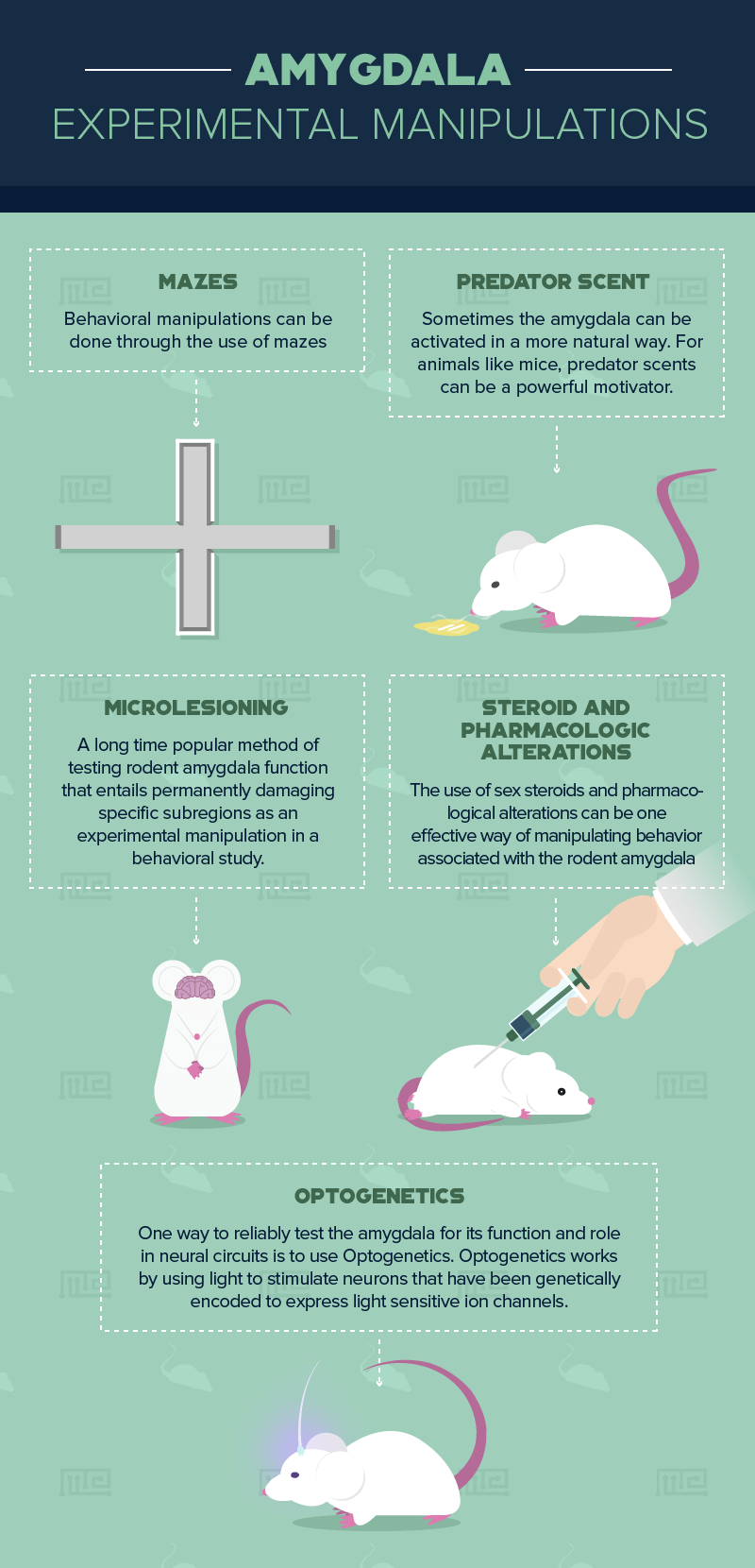
The rodent amygdala can be experimentally studied using behavioral, pharmacological, lesioning and genetic manipulations. The section below describes the various manipulations that can be achieved, first starting with behavioral manipulation via a maze, and next with chemical and neurobiological manipulations. The manipulations below include predator scent, pharmacologic or chemical agents that target amygdala subregion functioning, brain region lesioning of subregions of the amygdala to remove and inhibit functions connected to that subregion, and optogenetics in rodents genetically bred to have light-sensitive neurons that can be turned on and off with fiber optic light.
One important note for experiments with rodent or any animals; all animals should be handled in a consistent manner according to a study design protocol so that no variations in handling are introduced as confounding factors into the experiment. The conditions including the lab and environmental conditions cannot differ between tested animals. This is what is meant by controlling all other factors in an experiment. The experimental manipulation should be the only factor that changes.
Amygdala in Maze Learning
Vazdarjanova and McGaugh fear conditioned mice and then tested them in an elevated plus maze.[5] Their results revealed that the ventromedial prefrontal cortex forms a projection with the basomedial amygdala and that this pathway is essential for the suppression of freezing in fear-related states.[5] Mice treated and tested for spatial memory performance and fear extinction in a Y-maze allowed researchers to show that pharmacological inhibition of a glucocorticoid amplifying enzyme, highly expressed in limbic regions including the amygdala, can reverse the loss of spatial memory, and reduce contextual fear memory.[6]
One way to test the amygdala’s role in behavior is to test for anxiety-related behavior using a specially designed maze like the elevated plus maze. The elevated plus maze features an open elevated alley that induces approach avoidance. Unlike other, experiments the elevated maze does not have to rely on the introduction of noxious stimuli like predator scents, food deprivation or electric shock. Instead, the elevated plus maze design itself is a manipulated variable that contrasts the rodent’s preference for dark closed spaces with their unconditioned fear of heights and open spaces.[7]
A video tracking system is usually the main method for detecting and recording rodent activity after it enters the open or closed maze arms, but that is usually paired with manual observations. Both the video tracking and manual lab assistant notes are used to record how long a rodent may spend in each arm of the maze.[7] For the purposes of the experiment, the maze itself is a manipulation where anxiety and delay are expected of the rodents when they are in the elevated and open spaces, which can then be easily compared to rodent behavior in closed and dark spaces. Though a well-designed maze can be a natural experiment since it has both face and construct validity as an experimental instrument, it can also be the staging ground for experiments with the amygdala and other limbic regions that mediate anxiety and fear
Predator Scent
Sometimes the amygdala can be activated in a more natural way. For animals like mice, predator scents can be a powerful motivator. Research has shown that mice stressed by predator scents in a learning task improved their performance in the Morris water maze.[8] Emotional arousal activation of the amygdala is an important moderator of long-term memory formation.[9] The behavioral implications of this are important and can range from translational work to help improve the cognition functioning and quality of life in special patient populations, while also contributing to basic research on fear conditioning.
Steroid and Pharmacologic Alterations
The amygdala as part of the limbic system contributes to inhibitory control of impulse behavior. The use of sex steroids and pharmacological alterations can be one effective way of manipulating behavior associated with the rodent amygdala and the role it plays in regulating impulse control for reward seeking behaviors. Masaki et al.[10] used parachloroamphetamine (PCA), a known neurotoxin for 5-HT receptors, to test rodent behavior in Go/No Go and reversal learning tasks. The cued Go/No Go task measures impulse control and reversal learning is used to measure flexible behavior. Rodents were administered the PCA drug, trained on tasks in an operant chamber, observed in the Go/No Go tasks and then later sacrificed for brain dissection. The amygdala and other regions were dissected and tested for monoamine concentration. Analysis of the dissected amygdala showed a significant reduction in the concentration of 5-HT. Furthermore, the statistical analysis results showed a strong negative correlation between decreased 5-HT concentrations and rodent learning performance.[10]
Microlesioning
One long time popular method of testing rodent amygdala function entails lesioning, or permanently damaging specific subregions as an experimental manipulation in a behavioral study. The basolateral amygdala has been a popular target for behavioral studies. Another reason the basolateral amygdala makes a good target for manipulation is that it receives inputs from a variety of medial temporal lobe and cortical regions including the thalamus, hippocampus, orbitofrontal cortex and the medial prefrontal cortex[11][12] The basolateral amygdala plays a major role in monitoring and responding to sensory input linked to emotional valence, vigilance, and arousal.[11]
In a recent study on the role of the basolateral amygdala, Winstanley et al.[12] trained 32 male Lister Hooded rats to press levers to receive a food reward and to nose-poke in a specific area to trigger the levers to appear, and then trained in a delay-discounting task to study reward delay and impulse control. After all the rats were trained, 10 received lesioning of the basolateral amygdala, 12 received lesioning of the orbitofrontal cortex, and 10 received a vehicle infusion. They found that selective lesioning the basolateral amygdala increased impulsivity in the delay-discounting task. While the basolateral amygdala lesions resulted in apparent impulsivity of choosing rewards, the orbitofrontal cortex lesions decreased the impulsivity behavior. The lesion to the basolateral amygdala impaired that region in the rodent brain, and impaired its ability to link the memory that pressing the alternative B lever results in a large reward. However, the lesion did not alter rodent preference for a large reward in the absence of a delay; it merely impaired the memory of the value of the reward needed to guide behavioral choice.[12]
Optogenetics for Testing the Amygdala. (SHORTEN and review)
One way to reliably test the amygdala for its function and role in neural circuits is to use Optogenetics. Optogenetics works by using light to stimulate neurons that have been genetically encoded to express light sensitive ion channels. Channelrhodopsin-2 (ChR2) is an algal protein light-activated cation channel that is one of the main opsin discoveries responsible for a breakthrough in optogenetics.[13] The light-gated cation channel ChR2 is the most widely used optogenetic tool in neuroscience and neurobiology work. In order for ChR2 to work, its genetic expression must be altered and limited in specific groups of neurons. The three most common ways to change the gene expression include virus delivery, in vivo electroporation, and breeding transgenic animals.[13]
In an optogenetics experiment, regions of interest in the rodent brain are labeled with retrobeads, and then an optic wire is surgically placed into the subregion stereotaxic coordinates. In their 2015 work, Namburi et al.[14] virally targeted the nucleus accumbens and the centromedial amygdala to show that projections from the basolateral amygdala to those regions result in opposing synaptic changes that occur after fear or reward conditioning. They used optogenetics to effectively turn these regions on or off so that they could study the resulting effects. Namburi et al.[14] found that photostimulation of the nucleus accumbens resulted in positive reinforcement and that photostimulation of the centromedial amygdala mediated negative reinforcement by impairing fear conditioning and enhancing reward responses.
Optogenetics can be used in behavioral experiments where the animal is free moving, given that the fiber optic connection line is given sufficient length. This method allows researchers to use neuronal areas that can be triggered on and off with light, to better observe and quantify the neural correlates of emotional arousal and sensory behavior.[13] Optogenetics provides the advantage of allowing light sensitive neuronal regions to be altered non-destructively for experimentation. For more detailed information on optogenetics protocols visit the Deisseroth Lab’s optogenetics site hosted at Standford University.
Conclusion
Animal model experiments using a variety of genetic, chemical and lesion manipulations to the amygdala have revealed much about the function of amygdala subregions. Rodent experiments are used extensively as early-stage research for human behavior and in many cases are the staging grounds for future research in non-human primates and humans. Projections from the amygdala to cortical regions can be targeted in future research interested in translating knowledge of amygdala function into therapeutic interventions.
References
- Sokolowski, K., & Corbin, J. G. (2012). Wired for behaviors: from development to function of innate limbic system circuitry. Frontiers in molecular neuroscience, 5,
- Balderston, N. L., Schultz, D. H., Hopkins, L., & Helmstetter, F. J. (2015). Functionally distinct amygdala subregions identified using DTI and high-resolution fMRI. Social Cognitive and Affective Neuroscience, 10(12), 1615–1622. http://doi.org/10.1093/scan/nsv055
- Gafford, G. M., & Ressler, K. J. (2016). Mouse models of fear-related disorders: cell-type-specific manipulations in amygdala. Neuroscience, 321, 108-120.
- Adhikari, A., Lerner, T. N., Finkelstein, J., Pak, S., Jennings, J. H., Davidson, T. J., … & Kim, S. Y. (2015). Basomedial amygdala mediates top-down control of anxiety and fear. Nature, 527(7577), 179.
- Vazdarjanova, A., & McGaugh, J. L. (1998). Basolateral amygdala is not critical for cognitive memory of contextual fear conditioning. Proceedings of the National Academy of Sciences, 95(25), 15003-15007.
- Wheelan, N., Webster, S. P., Kenyon, C. J., Caughey, S., Walker, B. R., Holmes, M. C., … & Yau, J. L. (2015). Short-term inhibition of 11β-hydroxysteroid dehydrogenase type 1 reversibly improves spatial memory but persistently impairs contextual fear memory in aged mice. Neuropharmacology, 91, 71-76.
- Walf, A. A., & Frye, C. A. (2007). The use of the elevated plus maze as an assay of anxiety-related behavior in rodents. Nature protocols, 2(2), 322.
- Galliot, E., Levaillant, M., Beard, E., Millot, J. L., & Pourié, G. (2010). Enhancement of spatial learning by predator odor in mice: involvement of amygdala and hippocampus. Neurobiology of learning and memory, 93(2), 196-202.
- McGaugh, J. L., Cahill, L., & Roozendaal, B. (1996). Involvement of the amygdala in memory storage: interaction with other brain systems. Proceedings of the National Academy of Sciences, 93(24), 13508-13514.
- Masaki, D., Yokoyama, C., Kinoshita, S., Tsuchida, H., Nakatomi, Y., Yoshimoto, K., & Fukui, K. (2006). Relationship between limbic and cortical 5-HT neurotransmission and acquisition and reversal learning in a go/no-go task in rats. Psychopharmacology, 189(2), 249-258.
- Rosen, J. B., & Donley, M. P. (2006). Animal studies of amygdala function in fear and uncertainty: relevance to human research. Biological psychology, 73(1), 49-60.
- Winstanley, C. A., Theobald, D. E., Cardinal, R. N., & Robbins, T. W. (2004). Contrasting roles of basolateral amygdala and orbitofrontal cortex in impulsive choice. Journal of Neuroscience, 24(20), 4718-4722.
- Britt, J. P., McDevitt, R. A., & Bonci, A. (2012). Use of channelrhodopsin for activation of CNS neurons. Current protocols in neuroscience, 2-16.
- Namburi, P., Beyeler, A., Yorozu, S., Calhoon, G. G., Halbert, S. A., Wichmann, R., … & Gray, J. M. (2015). A circuit mechanism for differentiating positive and negative associations. Nature, 520(7549), 675.